MRI
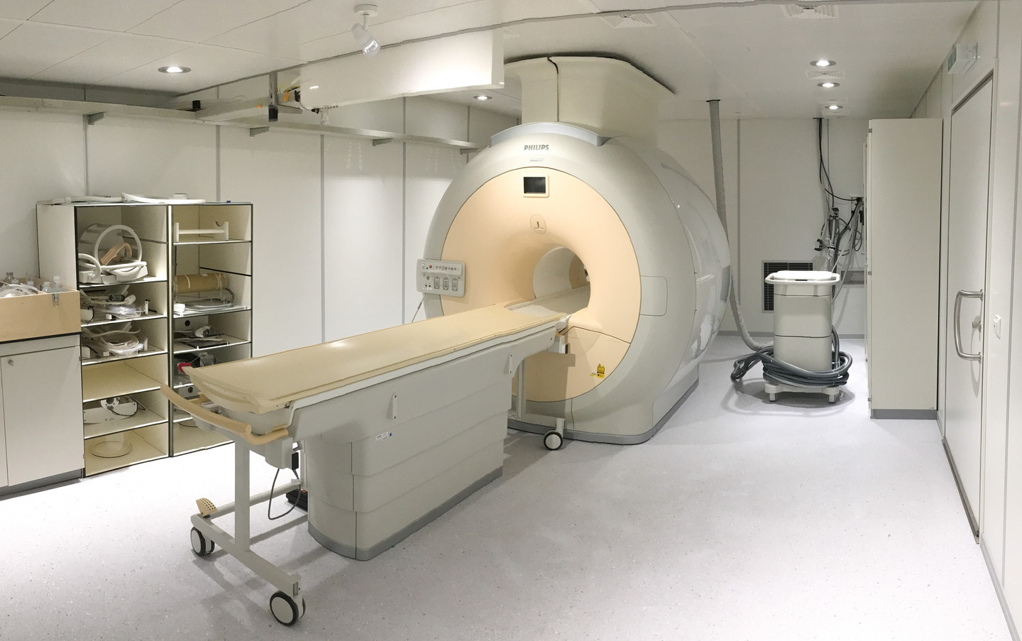
Magnetic Resonance Imaging (MRI) allows high resolution 3D imaging of soft tissue based on the magnetic properties of protons which are abundantly present in tissue. The object is placed in a strong magnetic field, which aligns a certain percentage of the magnetic nuclei of water protons along the field direction. The direction of the magnetized nuclei can be flipped using pulses of radiofrequency waves after which a relaxation towards the original direction occurs. As radiofrequency waves are emitted during the relaxation, the process of relaxation and its characteristic time can be measured. For image generation, MRI exploits the fact that relaxation times differ for different tissues, which allows to generate a plethora of specific images that weigh different properties in a specific manner. Thus, MRI can generate pictures that reflect the proton density, different relaxation properties, protons in fatty tissue, but also diffusion of water. Operating at a field strength of 3 Tesla, the current MRI (Achieva 3T, Philips Healthcare) allows to acquire images with an isotropic resolution of 250 µm. Image acquisition time depends on resolution, field of view and signal strength. The system can be used for all in vivo characterization of small and large animal models, especially to track tumor development in a longitudinal setting as no ionizing radiation is used.
Furthermore, the system is cabable to also image other elements with a suitable gyromagnetic ratio, for example Fluorine-19. As there is no F-19 present in soft tissue, fluorinated compounds can be used as contrast agents in applications related to image guided drug delivery.